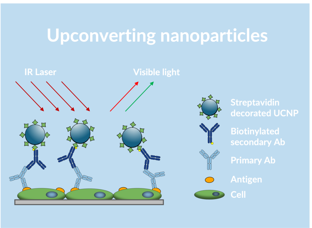
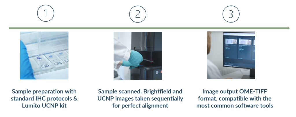
1. Tissue samples are prepared using standard IHC protocols
2. Streptavidin-decorated UCNPs bind to biotinylated primary or secondary antibodies supplied by the user
3. Near-infrared light excites the UCNPs
4. The UCNPs emit higher energy visible light
5. The scanner detects the emitted visible light without any autofluorescence or other tissue background
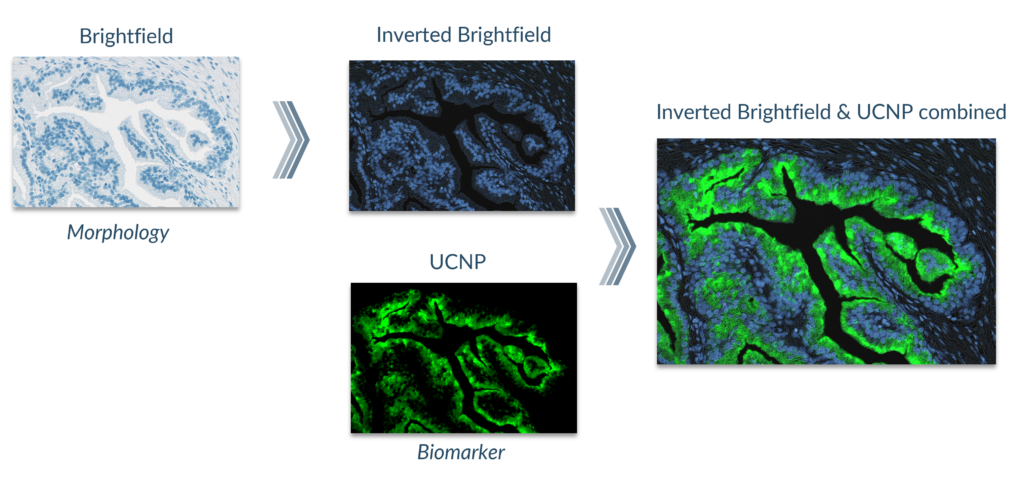
6. A separate brightfield image is acquired in series with the UCNP detection
Work in daylight
Easy to ship
Long storage (years)
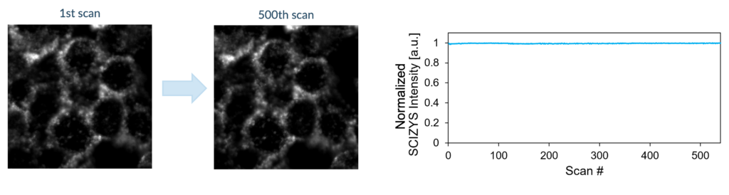
Rescannable slides