We have developed the next-generation IHC tissue analysis system, based on upconverting nanoparticles (UCNPs). The Lumito SCIZYS is a cutting-edge, high-sensitivity system combining labeling and imaging technologies to locate and measure biomarkers in tissue samples.
Overcoming limitations in tissue biomarker detection
Struggling to quantify low‑abundance biomarkers in tissue? SCIZYS enables ultra‑sensitive, autofluorescence‑free detection, with a wide dynamic range in a single image. Move beyond visual scoring.
Ultra-sensitive detection
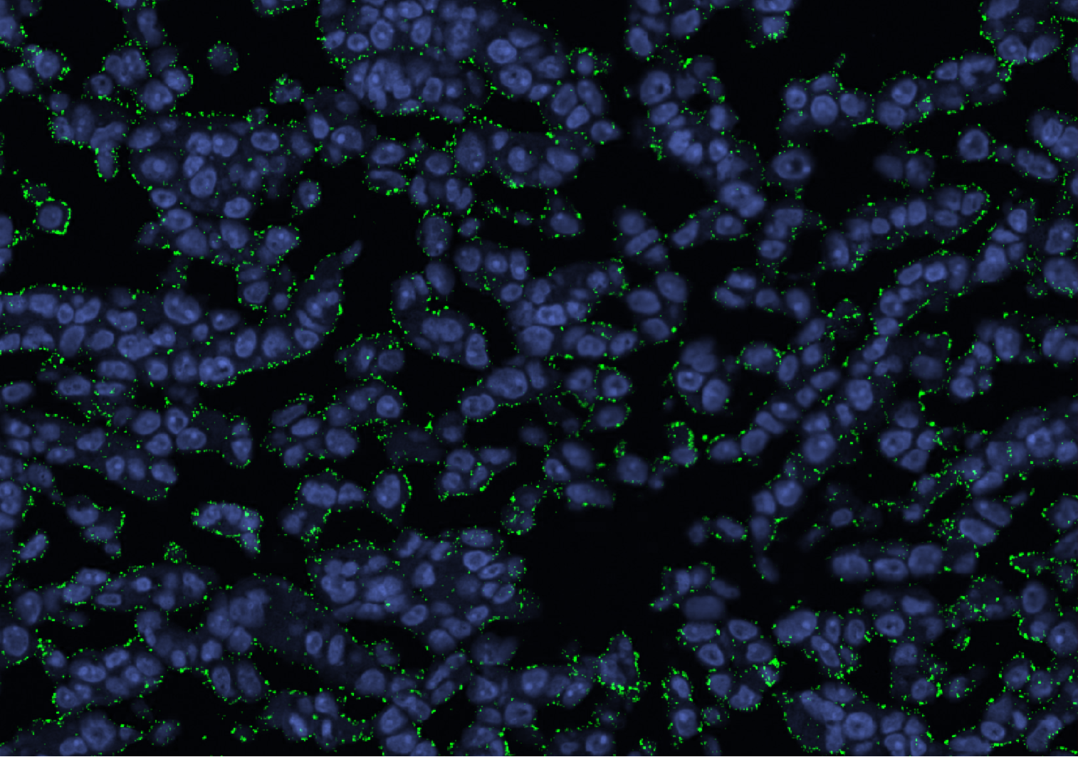
Detection down to single labels using photon-upconverting nanoparticles
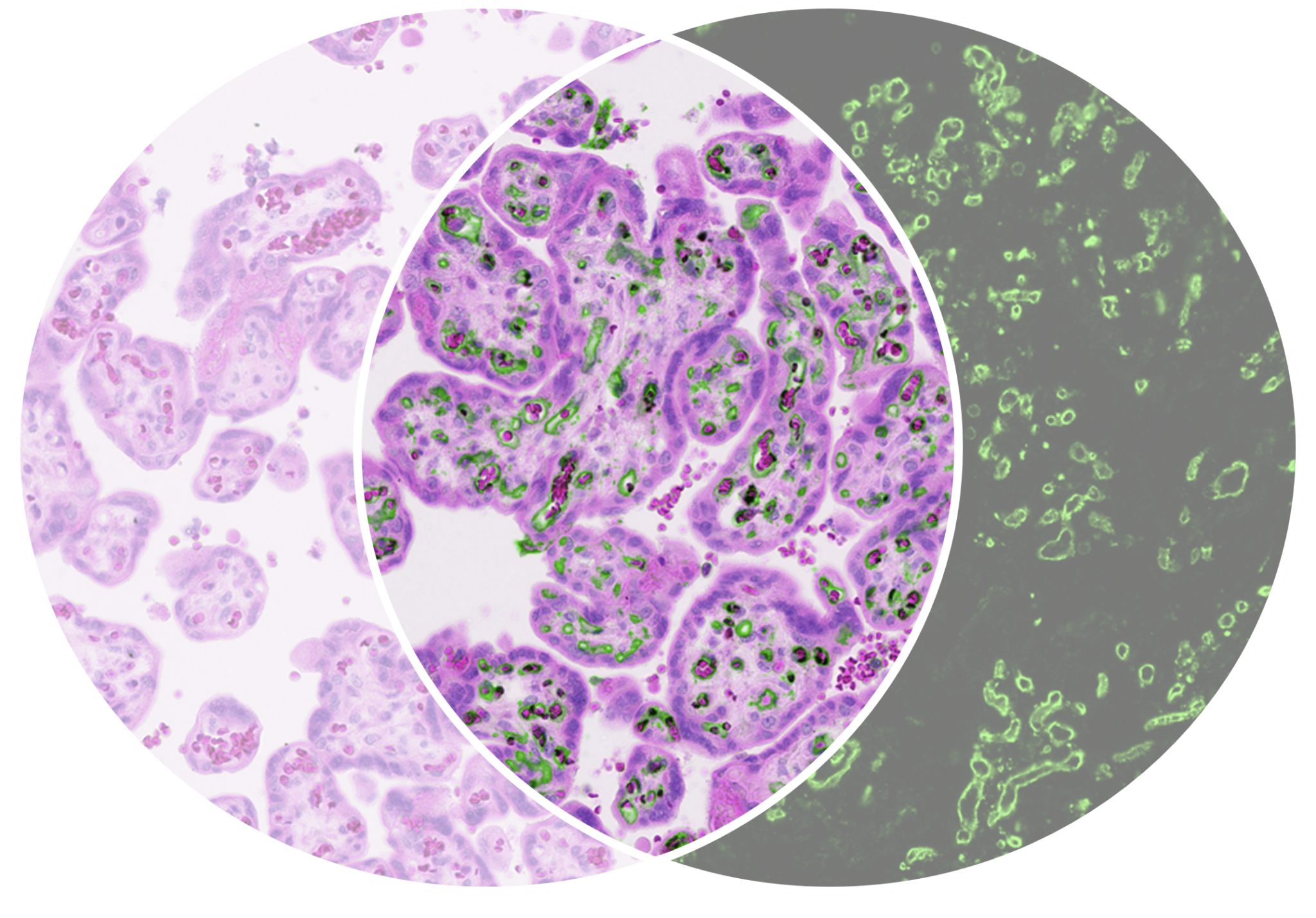
Full biomarker and morphology separation
Chromogenic counterstain and biomarker information in fully separated channels from the same sample
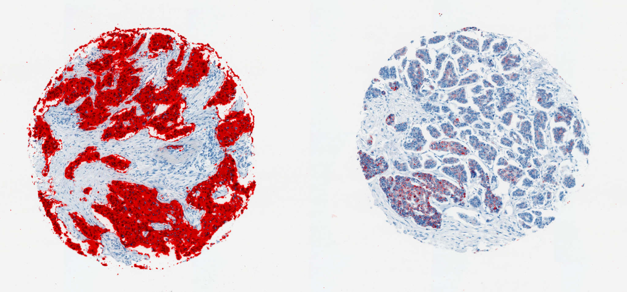
Exceptional dynamic range
High-accuracy quantification based on uniform nanoparticle light quanta and a wide dynamic range detection system
Scientific validation
Ultrasensitive HER2‑low detection
Measuring very low HER2 can be challenging with standard tissue methods. In our recent white paper we show how SCIZYS delivers up to 12× higher sensitivity and a broader dynamic range than traditional methods. We show how we can measure HER2 at levels that enable differentiation between HER2‑low and HER2‑negative tissue.
Detecting low-abundance biomarkers in tissue can be difficult with conventional methods. SCIZYS uses photon-upconversion nanoparticles (UCNPs) to eliminate tissue autofluorescence, achieve single-particle sensitivity, and provide exceptional photostability. The system enables precise detection of biomarkers like HER2 and PD-L1 at very low levels, offering a broader dynamic range and highly sensitive quantification for IHC applications.
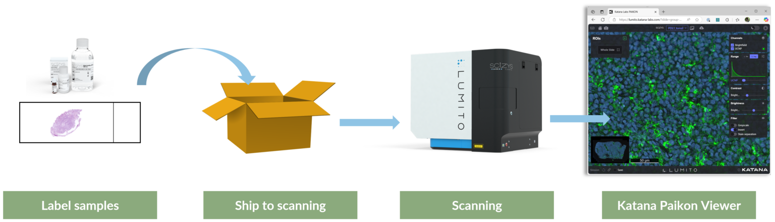
Lumito provides a complete workflow solution for ultra-sensitive tissue biomarker detection — from sample preparation to cloud-based image analysis.
1. Prepare IHC slides Use standard immunohistochemistry protocols.
2. Add SCIZYS detection reagents
Uses upconverting nanoparticle (UCNP) technology
Enables identification of biotinylated primary or secondary antibodies
Improves detection of biological markers in tissue samples
3. Send IHC slides to us for scanning Images histological samples using:
Brightfield imaging
UCNP luminescence detection
4. Access and analyze data You get access to the images in a browser based viewer, images can be analyzed directly or downloaded in standard ome-tiff format for further off-line analysis.
Research Use Only (RUO). Not for clinical or diagnostic use.
Request a technical discussion
For research teams interested in evaluating the platform.