
Lumito's technology makes it possible to stain tissue with nanoparticle-based reagents, and thus to create images with the company’s proprietary analysis instrument. Two of the major advantages are the ability to create imaging with high contrast and detail, and the ability to use both Lumito's reagent and traditional reagents to stain the same tissue section.
The images show a degree of detail comparable to the one achieved with traditional staining methods. The advantage of the imaging created with Lumito's technology is that only the signals from the intended markers are present – in this case, the breast cancer marker Her2.
– When you examine the Her2 imaging based on Lumito's technology, you can readily understand the opportunities it opens for pathologists when they are able to see only the tissue they are interested in. The technology further opens the door for image processing, which may help facilitate automated diagnostics in the future, says Stefan Nilsson, CEO of Lumito.
Lumito is also publishing cell imagery where UCNP staining and traditional H&E staining have been combined in the same section. The user may then choose to perform analysis on the images separately, or on the aggregate image
Images and captions:

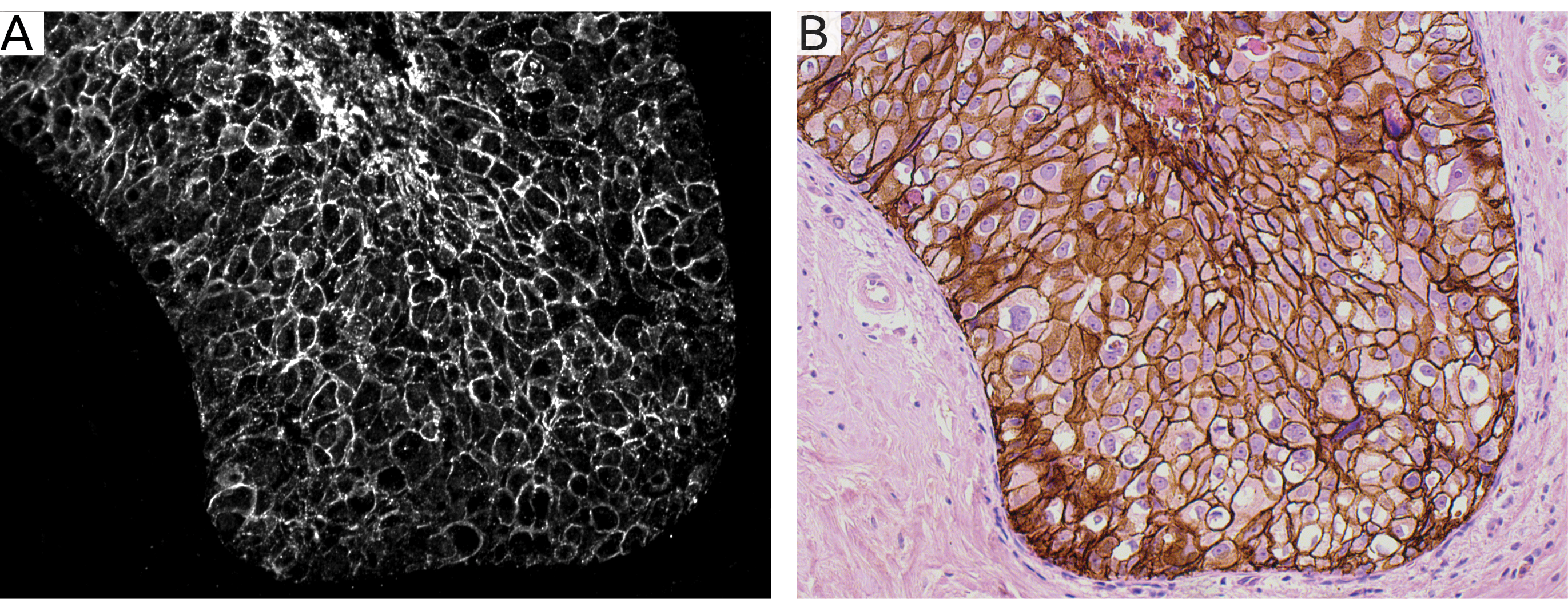
Figure 1. Comparison between Lumito's UCNP staining and the standard immunohistochemical DAB-based staining. In image A, the breast tissue has been stained with Her2-UCNP reagent, in order to visualize cells containing Her2, which is a common marker in breast cancer diagnostics.
Image B shows staining in the same tissue sample as in image A, but performed with the standard DAB-based method. Images A and B show a comparable level of detail, but both background signal and autofluorescence are eliminated in Lumito's UCNP-based staining. Eliminating all noise from background signal and autofluorescence improves the conditions for automated analysis based on for example AI algorithms significantly.

Figure 2. Combined UCNP and H&E staining in the same cell section.
Lumito's technology supports the performance of both UCNP and H&E staining in the same tissue sample. The user may then choose to perform analysis on the images separately, or on the aggregate image. Image A shows a UCNP-based Her2 stain of BT 474, a breast cancer cell line. In image B, the same section is shown with a standard H&E stain. Image C shows the aggregate image. The user may choose to perform analysis on both images separately, or, to improve accuracy and diagnostic reliability, on the aggregate image as shown here. (The identified cancer cells are visible in yellow).
Images are available for download: https://news.cision.com/se/lumito